
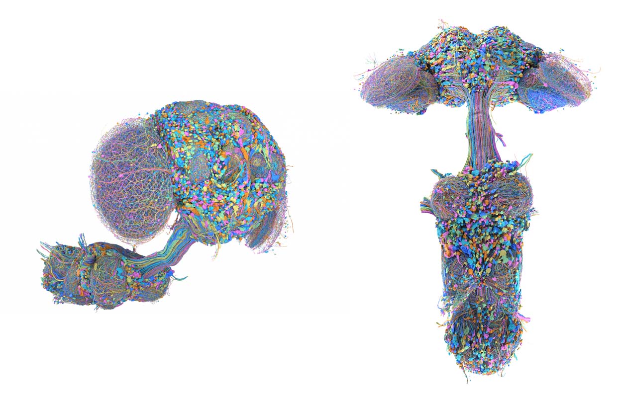
Inside a male fruit fly only a few millimetres long sits a central nervous system with more than 166,000 neurons and around 125 million synaptic connections. Google Research, working with HHMI Janelia and collaborators, has now mapped all those neurons and the connections between them. The result is the first complete connectome of the male fruit fly central nervous system and, when counting by neurons, the largest complete brain map to date.
Google has been working on connectomics for over a decade, using machine learning to help reconstruct increasingly large neural datasets. The work has moved from partial fly brains to human cortical tissue and now to complete nervous systems. Outside neuroscientific research, connectomics is also starting to find practical uses in neurosurgical planning, stimulation targeting, and computational models of brain function.
Google's map is not the first complete connectome map of the fruit fly brain. In 2024, the FlyWire consortium published a connectome of the adult female brain containing around 139,000 neurons and 54.5 million synapses. The new dataset extends that work across the entire male central nervous system, including the brain, optic lobes, and ventral nerve cord.
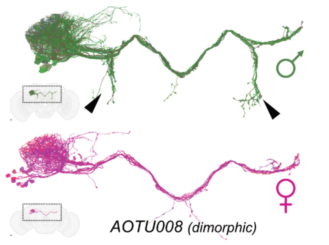
That makes it possible to compare complete male and female wiring diagrams. Researchers identified more than 100 sexually dimorphic cell types, alongside hundreds that appear only in one sex. Most sensory and motor circuitry was broadly shared. The clearest differences appeared higher up in the brain, including circuits involved in courtship and other sex-specific behaviours.
For Google, the project sits inside a coordinated research effort. Its dedicated Connectomics team was formed in 2014, with a focus on using machine learning and large-scale computing to reconstruct neurons from electron microscopy. In 2020, Google and Janelia released the fly “hemibrain”, mapping around 25,000 neurons and more than 20 million connections across part of the brain.
The scale has since moved beyond flies. In 2024, Google and Harvard reconstructed a cubic millimetre of human temporal cortex containing around 16,000 neurons and 150 million synapses. That tiny piece of tissue generated 1.4 petabytes (i.e. 1400 terabytes) of data. Google is also part of a $33 million, five-year effort to map 10-15 cubic millimetres of the mouse hippocampal formation; a dataset expected to reach roughly 25 petabytes.
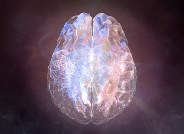
Connectomic mapping also has applications in humans, although at a very different scale from Google's fly work. Rather than resolving individual neurons and synapses, clinical approaches typically use diffusion MRI and functional MRI to map connections between larger brain regions and white-matter pathways. These network-level maps are used to understand how disease and intervention affect distributed brain circuits.
Omniscient Neurotechnology is among the frontrunners of applied connectomics. Its FDA-cleared Quicktome platform combines diffusion MRI and resting-state fMRI to generate patient-specific maps of brain networks and white-matter tracts that can assist neurosurgical planning. Omniscient raised $27.2 million earlier this year to expand the platform into areas including stroke, movement disorders, and BCI.
Connectomics is also being explored in brain stimulation. In connectomic deep brain stimulation, researchers look at the wider network connected to an electrode location instead of looking only at a target's anatomical coordinates. The approach is being studied across conditions including Parkinson's disease, depression, and OCD, to identify more precise stimulation sites linked to the circuits underlying specific symptoms.
Produced with support from

%20copy.jpg)





.svg)

