
Of the senses through which we experience the world, vision is perhaps the most coveted. The eye, connected through millions of nerve fibres to the brain, gives us a window into the world. And losing that window can be profoundly isolating. So when Neuralink founder Elon Musk declared that “we will cure blindness,” the promise quickly travelled across the world.
Vision, however, depends on a remarkably complex chain of events. Light must enter the eye, reach the retina, and be converted into electrical signals that travel through the optic nerve and deeper visual pathways before the brain interprets them. Problems can emerge at any point along the chain. Many vision complications are corrected at the start of the chain. But once the pathways carrying information toward the brain begin to fail, the problem becomes more complex.
This is where neurotechnology is pushing into new territory. New retinal implants can bypass damaged photoreceptors and feed information into surviving retinal circuits. Other systems aim to stimulate the retina without an implant. Cortical implants skip the eye entirely and write information directly into the brain. Neurotech researchers are approaching vision restoration from several points along the same pathway. But can they truly cure blindness?
Vision starts when light enters the eye and is focused onto the retina. Many common vision problems occur before light reaches the retina. For example, glasses correct how light is focused onto the retina, while cataract surgery can replace a clouded lens blocking light from passing through.
The retina is itself a layer of neural tissue. Rods and cones convert light into electrical signals, which pass through layered retinal circuitry before reaching retinal ganglion cells. Their axons come together to form the optic nerve. In conditions such as age-related macular degeneration and retinitis pigmentosa, photoreceptors progressively degenerate while parts of this downstream circuitry can remain intact.
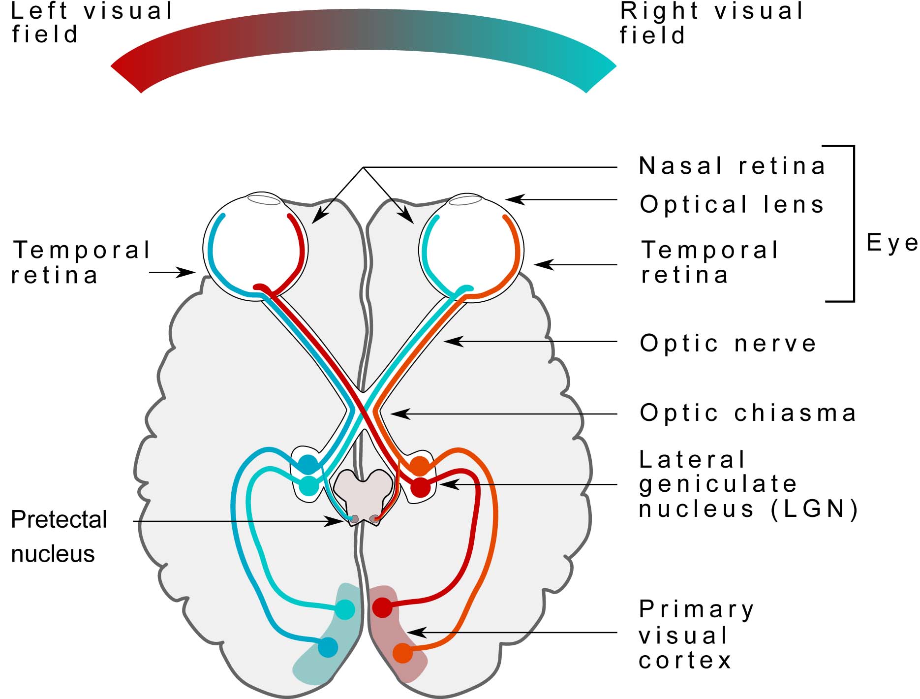
The optic nerve carries visual information into the brain. Much of the signal passes through the lateral geniculate nucleus, or LGN, before reaching the primary visual cortex, V1. This pathway retains a spatial organization of the visual field. But V1 is only the beginning of cortical visual processing. The signal then passes to further regions that interpret features like motion, depth, form, and the identity of what we see.
Where and how the visual pathway breaks determines the course of action. A patient with damaged photoreceptors may still have a functioning inner retina and optic nerve, making retinal stimulation possible. Damage to the optic nerve removes that route, pushing researchers further downstream toward the LGN or visual cortex.
Neurotech is already actively intervening at the retina.
Geographic atrophy, an advanced form of age-related macular degeneration, progressively destroys photoreceptors and supporting cells in the macula. As the affected area expands, patients lose the central vision used for reading, recognising faces, and seeing fine detail–even while some peripheral vision remains. Existing treatments slow progression in some patients, but they cannot restore photoreceptors that have already been lost.
Retinal prostheses replace that missing input. The main technology is Science’s PRIMA, a 2 by 2 millimetre photovoltaic chip implanted beneath the retina in the area of atrophy. Its 378 microscopic pixels act as artificial photoreceptors, converting incoming energy into electrical stimulation of surviving bipolar cells. From there, the patient's intact retinal circuitry and optic nerve carry the signal towards the brain.
The implant works with a pair of camera-equipped glasses. Images captured by the camera are projected onto PRIMA using near-infrared light, which simultaneously powers the implant and carries the visual pattern. Patients can adjust contrast and magnify parts of the image before they reach the retina. Because the system targets the damaged centre of the retina, the prosthetic image can be used alongside remaining peripheral vision.
The technology originated in Daniel Palanker's lab at Stanford before being developed commercially by French neurotech company Pixium Vision. Pixium took PRIMA into human studies and launched the pivotal PRIMAvera trial, but entered liquidation in 2024.
Science acquired the technology that year and continued its clinical and regulatory development. In 2026, PRIMA received a CE mark and entered commercial use in Europe, while Science continues to pursue FDA approval in the US.
In the pivotal study, 38 people with severe central vision loss received PRIMA. At 12 months, 26 of 32 evaluable participants achieved a clinically meaningful improvement in visual acuity, while natural peripheral vision remained stable. The latest 36-month follow-up showed improvements continued long after the initial procedure.

PRIMA is showing strong early results, but retinal implants still come with trade-offs. They require surgery, permanently place hardware inside the eye, and face limits on how precisely electrical current can stimulate neighbouring neurons.
Paris-based Axorus started from a similar premise, developing an electrical retinal implant after its founding in 2019. But the team eventually became concerned about how far electrical stimulation could scale, both in the precision of individual stimulation points and in the area of vision that could be covered.
The latter is particularly important. “If you have very high resolution, meaning high acuity, but the field of view is narrow, it’s like looking through a keyhole,” co-founder Jean-Damien Louise explains.
Axorus began searching for another way to stimulate surviving retinal cells. The team screened around 40 technologies before finding research from Boston University on photoacoustic stimulation. The technique offered a way to activate smaller groups of neurons without relying on densely packed electrodes. Axorus licensed the technology and initially developed a photoacoustic film implanted behind the retina.
“The photoacoustic effect looks like magic, but it isn’t,” Louise says. A pulsed laser hits a light-absorbing material, causing it to rapidly expand and contract and generate ultrasound waves. By shaping the projected light, Axorus can effectively draw an ultrasonic stimulation pattern across the retina.
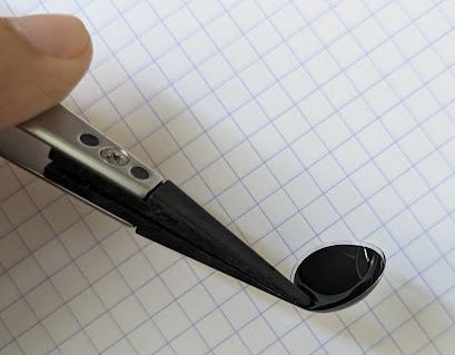
Axorus then found that the photoacoustic material did not need to sit inside the eye at all. Its current system places it in a contact lens, paired with camera-equipped glasses that capture the surroundings and project the corresponding pattern onto the lens. “We simply bought black contact lenses online, and it worked,” Louise says.
Axorus is initially targeting dry AMD and retinitis pigmentosa, but the approach remains preclinical. Animal experiments have produced retinal and downstream brain responses consistent with visual stimulation. The company is preparing for a first human study in 2027.
Retinal approaches assume enough of the eye and optic nerve is intact to carry information onwards. For people whose optic nerve is severely damaged, that route is closed. Cortical prostheses move further downstream, bypassing the eye entirely and stimulating the visual cortex at the back of the brain.
A camera captures the surroundings, software reduces that image into a stimulation pattern, and implanted electrodes attempt to turn it back into something the brain can interpret. As ambitious as it sounds, the principle is already being tested in humans.
Eduardo Fernández and the CORTIVIS group at Miguel Hernández University have implanted five blind volunteers with Utah arrays, inserting interfaces of around 100 electrodes in the primary visual cortex. Electrical stimulation then evokes phosphenes–artificial visual percepts that participants typically describe as points or shapes of light.
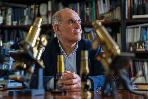
“Even if the perceptions are very simple, people can use them for mobility and orientation,” Fernández explains. “Sometimes they are even able to read some characters.”
Those percepts can differ substantially between people. Most participants reported black-and-white or yellowish sensations, but one volunteer could perceive several colours. Fernández does not know why. The participant had lost his sight around a year before implantation, more recently than the others, which may have played a role. “It could be related to that, but we do not have enough data to know.”.
The variation illustrates a deeper challenge: the visual cortex is not a screen. It is tempting to imagine every electrode adding a pixel to our vision, with higher electrode counts producing progressively sharper images. But Fernández says human experiments show something considerably messier.
One electrode might create a dot, while another produces a line, circle, or larger shape; percepts can also change between sessions. “Everybody says it is like pixels, and it is not like that,” he says. “One single electrode can produce a very complex perception.”
Fernández's group is studying closed-loop systems that record brain activity and adapt stimulation to the current state of the cortex, including the timing of pulses and interactions between electrodes. He points to cochlear implants as a useful precedent. Electrode counts have changed relatively little over decades, but better encoding strategies have dramatically improved what users can understand.
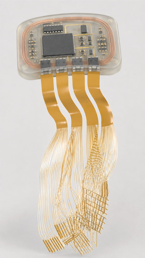
Belgian startup ReVision Implant is among the companies pushing the hardware towards patients. Founded in 2020, the company is developing thin, flexible electrode arrays designed for chronic stimulation of the visual cortex.
Camera-equipped glasses capture the environment and track where the wearer is looking, while software reduces the image before wirelessly sending stimulation instructions to the implant. The system is designed to reach different depths of the visual cortex, giving it access to both central and peripheral vision for a wider, more natural field of view, and will eventually stimulate around a thousand locations.
Rather than reproducing everything a camera sees, ReVision expects early systems to preserve only the most useful information. Contours, contrast, key features that help a user navigate their surroundings. “With a thousand pixels, if you use them correctly, you can already give a lot back to someone who sees nothing,” says CSO Laurens Goyvaerts.
Human testing is now approaching. ReVision has received FDA Breakthrough Device designation and is moving through a staged clinical roadmap. Its first step is an acute insertion study in patients already undergoing brain surgery, designed to show that the flexible array can safely be implanted in the human cortex.
A later experiment will place the visual prosthesis in a blind participant for several months, before progressing toward a chronic wireless system.
Neuralink is pursuing the same target at a much greater scale. Its Blindsight program would use robotically inserted flexible threads to stimulate the visual cortex, but the company has not yet tested visual stimulation in humans. “The robot they are using is really amazing,” Fernández says. “But we have not seen human stimulation data yet.”
Ultimately, cortical vision may depend as much on learning as on resolution. People who previously had sight are currently the clearest candidates because their visual system developed with normal input, while congenital blindness raises harder questions around how the cortex can reorganise to accommodate new input.
Even for previously sighted patients, Fernández does not expect an implant simply to recreate the original image. A user may instead learn that one artificial pattern signals a doorway and another an obstacle. Cortical prostheses therefore bypass more damaged biology than retinal approaches, but in doing so take on the harder task of teaching the brain a new visual language.
Between the retina and visual cortex sits another potential target. Dutch startup Phosphoenix is developing an implant for the lateral geniculate nucleus, or LGN, the thalamic structure that relays visual information towards the cortex.
The company grew out of work led by neuroscientist Pieter Roelfsema, whose group previously demonstrated high-channel-count stimulation of the visual cortex. Phosphoenix has since shifted its focus deeper into the visual pathway, aiming to bypass both the eye and optic nerve while stimulating the system before information reaches the cortex.
Its system combines camera-equipped glasses with the Fountain Probe, a flexible implant containing more than 1,000 stimulation electrodes. The probe will distribute those electrodes through the small volume of the LGN, where electrical stimulation would generate phosphenes that the brain could learn to interpret as visual information.
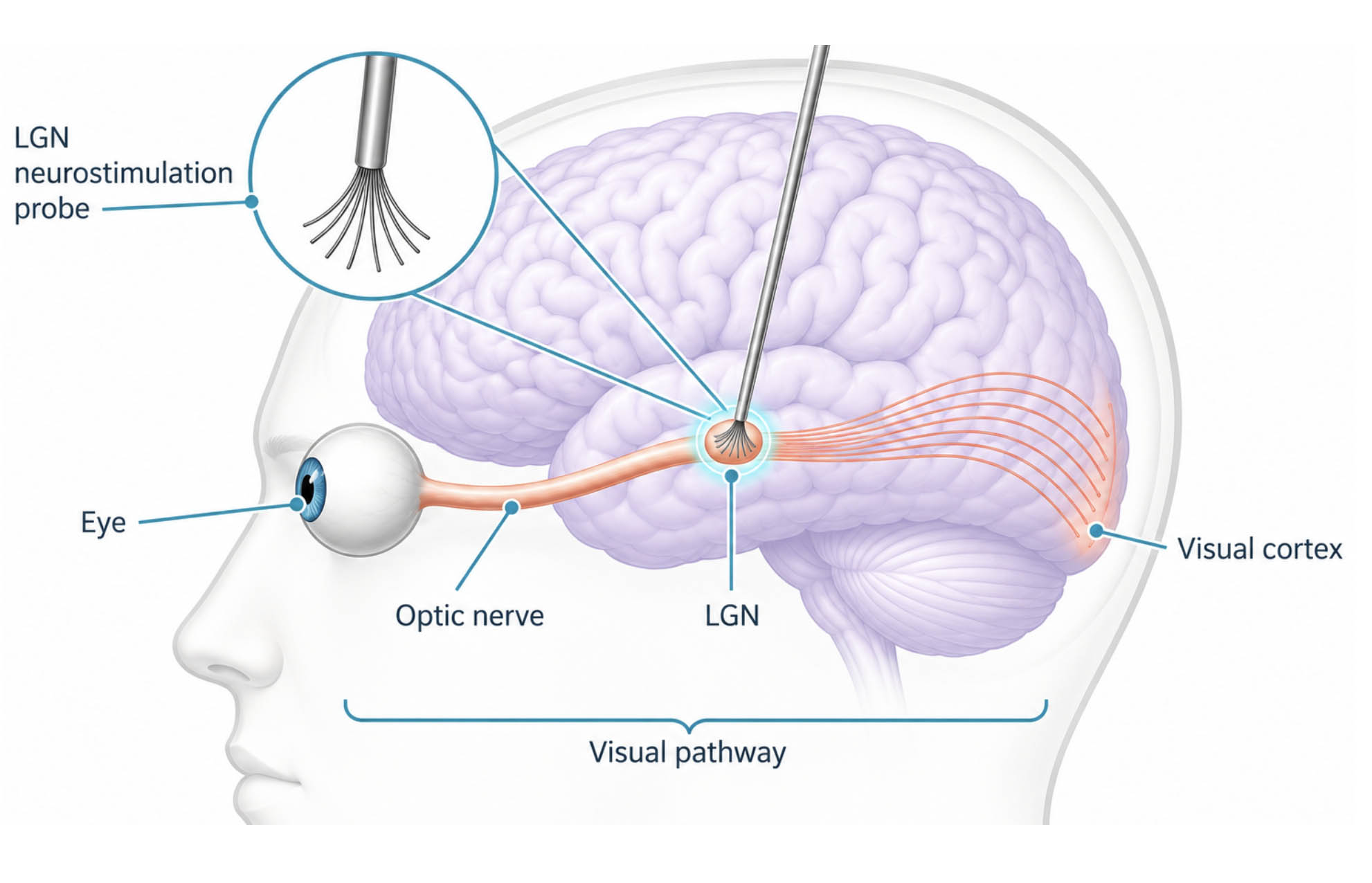
Phosphoenix believes that this compact relay structure could offer broad visual-field coverage without requiring electrodes to be spread across the much larger visual cortex, although that still needs to be demonstrated clinically.
Phosphoenix is now moving towards that test. In September 2026, the company raised €1.3 million to prepare for its first-in-human study and further develop the stimulation system, alongside a €2.5 million EIC Transition grant supporting the wider SIGHTED project. The current plan is to implant five blind participants in a first clinical study targeted for 2027.
And of course, hardware is only one route to restoring lost visual input. For example, a group of biotech companies is now trying to make surviving retinal cells light-sensitive through optogenetic gene therapies.
There is unlikely to be a single route to restoring vision. Progress will depend on matching the intervention to where the visual pathway has failed, from the retina to the LGN and ultimately the cortex. But for many visually impaired people, neurotechnology could one day restore part of the vision they have lost.
Produced with support from



%20copy.jpg)


%20copy.jpg)

.svg)

